
Mole Removal Singapore
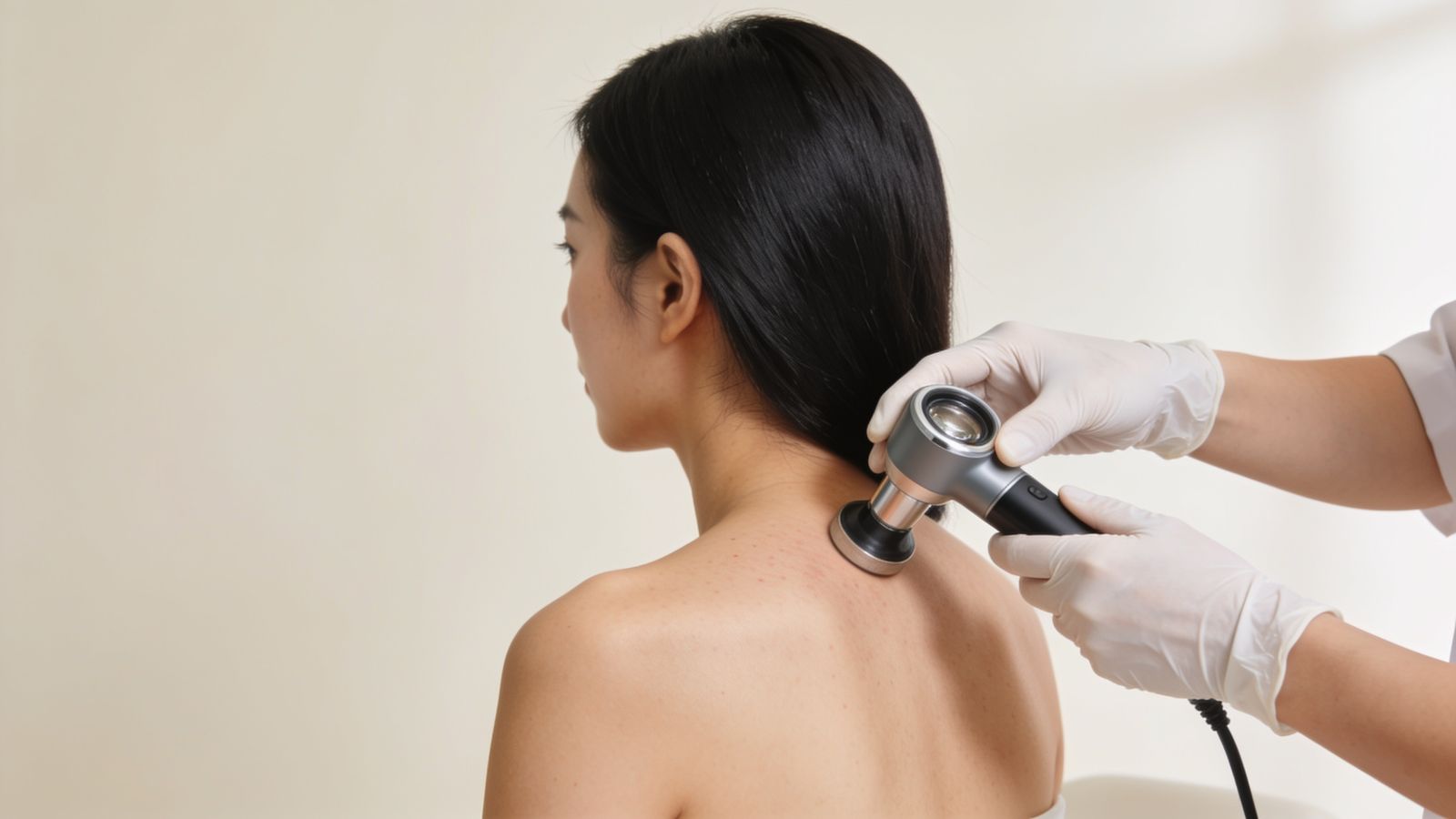
Before any mole is removed, it is examined with a dermatoscope. At IN Eternity Clinic, anything suspicious is referred for specialist assessment rather than removed for appearance.
WhatsApp to Enquire →Key facts ↓Mole removal in Singapore is a minor procedure that takes off a benign mole by CO2 laser, shave or excision; at IN Eternity Clinic every mole is dermatoscope-checked first. Clearly benign raised moles may be shaved or lasered level with the skin; flat, deep or doubtful moles suit excision, which sends the whole mole to the laboratory. Anything suspicious is referred for specialist assessment.
- Sessions: Mole removal is usually one visit, and several small benign moles can often be treated in the same session.
- Recovery: After laser or shave, a small scab lifts over one to two weeks; after excision, stitches are removed at around one to two weeks.
- Comfort: A small local anaesthetic injection stings for a few seconds; after that, removal is felt as pressure only.
- Cost: Depends on the number, size and site of the moles, the method and any laboratory examination; the S$120 consultation fee is waived with any medical treatment.
- Who treats you: Dr Sin Yong examines every mole with a dermatoscope and refers anything suspicious rather than removing it for appearance.
Comparing methods? See mole removal: laser vs shave vs excision.
Mole removal in Singapore at IN Eternity starts with a dermatoscope check; a mole that looks suspicious is referred for biopsy rather than lasered.
- It is new in adulthood, or changing in size, shape or colour.
- It bleeds, itches or crusts without injury.
- It has more than one colour, an uneven edge, or looks unlike your other moles (the “ugly duckling” sign).
- It sits on the palm, sole, nail or genital skin.
These moles are referred for specialist assessment and, where needed, excision with laboratory examination, not lasered for looks. See mole check before removal.
Mole removal at a glance
| What it is | A minor procedure to remove a benign mole by CO2 laser, shave or excision, after a dermatoscope check. |
|---|---|
| Used for | Clearly benign raised or flat moles removed for appearance; anything suspicious is referred for specialist assessment. |
| How it works | Raised benign moles are shaved or lasered level with the skin; flat, deep or doubtful moles suit excision, which sends the whole mole to the laboratory. |
| Typical sessions | Usually one visit; several small benign moles can often be treated in one session. |
| Downtime | A small scab that lifts over one to two weeks after laser or shave; stitches after excision, removed at around one to two weeks. |
| When results show | The pink mark fades over months. |
| Who should avoid or wait | Moles that are new, changing, bleeding or atypical are not removed for appearance; keloid-prone sites such as the chest, shoulders, upper back and jawline need extra caution. |
| Who performs it | Medical Director Dr Sin Yong examines every mole with a dermatoscope and chooses the method. |
Dr Sin Yong, Medical Director of IN Eternity Clinic, explains that laser leaves nothing to examine, so every mole is scoped first and anything doubtful is excised for the laboratory or referred, rather than lasered for appearance.

Shave, CO2 laser or excision?
| Method | How it works | Usually suits | Downtime | Scar | Laboratory check |
|---|---|---|---|---|---|
| Shave removal | A fine blade takes the mole level with the skin under local anaesthetic | Raised moles | Small crust for about a week | Flat pale mark; may fade | Yes, the tissue can be sent |
| CO2 laser ablation | Vaporises the mole in thin layers | Small flat or slightly raised moles judged benign | Crust for about a week | Small pale or pink mark | No; tissue is destroyed, so only for moles cleared on dermatoscopy |
| Excision with stitches | Cuts the mole out with a margin and closes the skin | Deep moles, large moles, any mole that needs full examination | Stitches for one to two weeks | Fine line scar | Yes, always |
On this page
Consultations in English and Mandarin (面诊可使用英语或华语). This page is also available in: 中文.
- First step: dermatoscopic examination of the mole and its history: new, changing, bleeding or itchy.
- Warning signs (ABCDE): asymmetry, irregular border, uneven colour, diameter over about 6 mm, and evolution over time.
- If anything is suspicious: referral for specialist assessment and, where needed, excision with laboratory examination.
- Benign moles: removed by a method matched to type and depth, such as CO2 laser shaving or excision.
- Scar: every method leaves a mark of some kind; size, site and skin type are discussed before choosing.
- Where: IN Eternity Clinic, 9 Scotts Road, #12-01 Pacific Plaza, Singapore.
- Why the dermatoscope comes first: a meta-analysis of nine studies found the diagnostic odds for melanoma were 15.6 times higher with dermoscopy than with the naked eye (9.0 after two outlier studies were removed). (Source: Vestergaard 2008) [1]
- Shave removal: of 204 common moles removed by shave excision, the result was rated excellent in about a third and poor in about 1 in 12, and the mole returned, clinically or under the dermatoscope, in 40 (about 1 in 5). (Source: Ferrandiz 2005) [4]
- Excision vs laser: pooling 46 studies and 4,201 moles, surgical excision cleared 96 in 100 with the lowest recurrence (about 2 in 100); after CO2 laser about 13 in 100 recurred. (Source: Guo 2026)
- Appearance: in the same analysis cosmetic satisfaction scored higher after laser (8.4 out of 10 for CO2) than after excision (6.2), and deeper intradermal moles were about 1.8 times as likely to recur as flat junctional ones. (Source: Guo 2026)
Why the dermatoscope comes first
A dermatoscope magnifies the mole and shows pigment patterns beneath the surface. Studies show it improves the accuracy of melanoma detection compared with the naked eye. A mole that looks harmless to the eye can show features that call for caution, and the reverse.
Removing a mole by laser vaporises the tissue, so nothing is left to examine under a microscope. That is why any doubt leads to referral and excision with laboratory examination rather than cosmetic removal.

Assessment decides the method
Flat, benign pigmented moles and raised soft moles behave differently. Raised benign moles are often shaved level with a CO2 laser on the DEKA platform, leaving a small mark that fades over months. Deeper moles may be better excised and stitched. Pigment that remains in the deeper dermis can recur, which is discussed beforehand.
Red spots that look like moles are often cherry angiomas, and rough, stuck-on brown spots are often seborrhoeic keratoses; both are handled differently.
“A mole removed by laser can never be examined under a microscope. That is why every one is scoped before anything is removed.”
When a mole should be checked
Any mole that is new in adulthood, changing in size, shape or colour, bleeding, itching or looking different from your other moles should be checked promptly, whether or not you want it removed.
Laser mole removal vs shave vs excision: which suits which mole?
The three methods differ in what they remove, whether anything can be sent to the laboratory, and the mark they leave. The choice follows the mole — its type, depth, site and how certain the diagnosis is — rather than preference for one technique.
| Method | Usually suits | Laboratory sample | Typical mark |
|---|---|---|---|
| CO2 laser | Small, raised moles that are clearly benign on dermatoscopy | No — tissue is vaporised | Small flat mark, pink then paler over months |
| Shave removal | Raised, soft, benign moles on the face or body | Yes, the shaved tissue | Flat mark; some pigment may return |
| Excision | Flat or deeper moles, and any mole with doubt | Yes, the whole mole with a margin | Fine line scar after stitches |
Laser and shave methods leave pigment cells in the deeper skin, so a mole can partly regrow, sometimes looking irregular. A regrown mole is always re-examined, and the original appearance helps interpret it, which is why photographs are taken before treatment. Excision is less likely to recur but leaves a line scar and is planned along natural skin creases.
ABCDE, the “ugly duckling” and EFG: when must a mole be checked before anything else?
The ABCDE signs are a prompt to get checked, not a diagnosis: Asymmetry, irregular Border, uneven Colour, Diameter over about 6 mm, and Evolution — any change in size, shape, colour or height. Of these, change over time is the most important. A mole that looks unlike your other moles, the “ugly duckling”, also deserves a closer look.

Some melanomas do not follow ABCDE. A new bump that is Elevated, Firm and Growing over weeks to months (EFG) should be seen promptly, even if it is pink or skin-coloured. At assessment, Dr Sin Yong examines the mole with a dermatoscope, compares it with your other moles and takes photographs. A mole with concerning features is not removed for appearance; you are referred to a dermatologist for excision and laboratory examination. A borderline mole may instead be photographed and re-examined after an interval to look for change.
Most moles are harmless, and the point of the check is to find the few that are not. Our guide to moles explains the common types and when to have one examined, whether or not you want it removed.
What affects the cost of mole removal?
Cost depends on the number of moles, their size and site, the method that suits each one, and whether a laboratory examination is needed. Removing several small benign moles in one session is planned differently from excising a single mole that needs a sample. For a breakdown of these factors, read the mole removal cost guide. Yellow eyelid plaques are a different condition from moles; xanthelasma removal has its own page.
If your spot may not be a mole at all, these pages may help: red spots are often cherry angiomas, rough brown patches are often seborrhoeic keratoses, and soft hanging lumps are usually skin tags. For the wider picture, see skin growths.
| Factor | Why it changes the cost |
|---|---|
| Number of moles | Several small benign moles in one session are planned differently from a single mole. |
| Size and depth | Larger or deeper moles take longer and are more likely to need excision. |
| Site | The face, eyelid margin and lip need finer work; moles on the palms, soles and nails are usually excised or referred. |
| Method | Laser, shave and excision differ in time, materials and dressings. |
| Laboratory examination | A sample sent for laboratory examination adds a laboratory fee. |
| Stitches and review | Excision includes stitch removal; a mole that regrows is re-examined. |
At IN Eternity Clinic a consultation fee applies, and it is waived if you go ahead with any medical treatment; the fee is S$120. See fees.
Published price ranges in Singapore. The table below summarises prices other Singapore clinics publish, as a guide to the market; it is not a quote. Your plan and cost are set at consultation (S$120, waived if you go ahead with treatment the same day).
| What is listed | Published price | Clinics | Sources |
|---|---|---|---|
| Mole or skin growth removal (laser or radiowave) | S$200–S$600 (per growth, per area or “from” prices) | 5 | Illumia Medical Aesthetics, David Loh Aesthetics, Lux Medical Aesthetic Clinic, A.M Aesthetics, SL Aesthetic Clinic read 20 Oct 2026 |
What does the dermatoscope check actually look for?
A dermatoscope is a hand-held magnifier with its own polarised light that cuts surface glare, so the doctor can see colour and structure a fraction of a millimetre below the surface. Ordinary moles tend to show an even, fine brown net, or evenly spaced round dots and globules, with symmetry from one side to the other. Features that call for caution include an irregular or broken net, streaks at the edge, blotches of uneven colour, blue-white or grey areas, scar-like white zones and unusual blood vessel patterns.
Site matters. Moles on the palms and soles have their own patterns that follow the skin ridges, and a pattern that sits on the ridges rather than in the furrows needs attention. A new brown or black streak in a nail should be examined. Facial moles in older skin can be hard to tell from flat sun spots. A dermatoscope improves accuracy, but it does not give a final diagnosis; only laboratory examination of removed tissue does that, which is why any doubt leads to excision rather than laser.
Which method suits which kind of mole?
The comparison above is organised by method. Patients usually start from the mole, so this table works the other way round.
| Mole or lookalike | How it usually looks | Method usually considered |
|---|---|---|
| Raised, soft, skin-coloured or light brown mole on the face | Dome-shaped, smooth, unchanged for years | Shave or CO2 laser if clearly benign on dermatoscopy |
| Flat brown mole | Even colour, level with the skin | Often left alone; excision if removal is wanted, as laser tends to leave pigment that returns |
| Mole present since birth or larger than usual | Larger, sometimes hairy | Planned excision or dermatology opinion, depending on size and site |
| Changing, irregular or itchy mole | Uneven colour or edge, or recent change | Referral for excision with laboratory examination |
| Rough, stuck-on brown spot | Waxy surface, often in sun-exposed skin | Usually a seborrhoeic keratosis, treated by laser or curettage |
| Blue-grey firm spot | Uniform blue colour | Usually excision so it can be examined |
Does mole removal hurt? Comfort and what to expect day by day
A small injection of local anaesthetic is given first, which stings for a few seconds; after that, laser, shave or excision is felt as pressure only. Day 0: a small dressed wound or scab. Days 1–3: keep it clean and dry as advised, with a thin layer of the ointment provided. Week 1: laser and shave sites form a scab that lifts on its own over one to two weeks; stitches, if used, are removed at around one to two weeks depending on the site. Weeks 2–4: the mark is pink and fades over months; sun protection reduces darkening. Review: the result and any laboratory report are discussed with you.
Mole removal aftercare and scarring: what to expect
Every method leaves a mark of some kind. What aftercare can do is keep it small and flat and stop it darkening. The first two weeks are about a clean, moist wound; the months after are about sun and time.
| Method | First week | Usually healed | What the mark tends to become |
|---|---|---|---|
| CO2 laser | Thin scab; ointment as advised; keep dry for the first day | Scab lifts in one to two weeks | A small pink spot that fades over months; may end slightly paler or darker |
| Shave removal | Shallow wound with a scab; ointment; light dressing where clothing rubs | One to two weeks | A flat mark; some pigment may return |
| Excision | Stitches; keep the dressing dry as advised; avoid stretching the area | Stitches out at about one to two weeks | A fine line that matures over six to twelve months |
- Let the scab come away on its own; picking raises the chance of a pale or dark spot.
- Use sunscreen or keep the site covered for at least three months, because new skin darkens easily in skin types III to V.
- Use silicone gel once the wound has closed, where advised, especially on the chest, shoulders and jawline.
Some sites scar more than others. The chest, shoulders, upper back and jawline are prone to raised or keloid scars, so moles there are planned with this in mind; a personal or family history of keloids changes the advice, and sometimes the honest answer is to leave a harmless mole alone. Contact the clinic if a site becomes increasingly red, hot, swollen or painful, if it bleeds again after the first day, or if pigment reappears. A regrowing mole is always re-examined. General wound care is set out on our aftercare page, and raised scars are covered under surgical and trauma scars.
Laser vs excision for moles: what the evidence shows about recurrence and scarring
A 2026 systematic review and meta-analysis pooled 46 studies covering 4,201 moles (melanocytic naevi) and compared the removal methods directly. Surgical excision gave the highest clearance and the lowest recurrence, at 2.1%. The non-surgical methods recurred more often: CO2 laser in 12.9% of cases, Er:YAG laser 14.3%, electrosurgery 16.1% and dermabrasion 22.7%.
| Method | Recurrence reported | Cosmetic satisfaction (out of 10) |
|---|---|---|
| Surgical excision | 2.1% | 6.2 |
| Shave excision | Not pooled in this analysis | 7.6 |
| CO2 laser | 12.9% | 8.4 |
| Er:YAG laser | 14.3% | 8.8 |
| Electrosurgery | 16.1% | – |
| Dermabrasion | 22.7% | – |
The cosmetic ranking runs the other way. That inversion is the whole decision in one line: the method that clears most reliably leaves the most visible mark, and the method that leaves the smallest mark is the one most likely to let the mole return.
Why depth decides whether a mole comes back
Junctional naevi sit at the junction between the epidermis and the dermis; intradermal naevi extend deeper. A laser vaporises tissue to a controlled depth, so naevus cells below that depth remain and pigment can return as they repopulate. In the same analysis, recurrence was significantly higher for intradermal than for junctional naevi (relative risk 1.82). That is not a failing of laser as a technology; it is a mismatch between a surface-limited method and a mole that is not surface-limited.
Looking properly while removing also changes the result. Dermatoscope-guided shave removal reported recurrence of 7.8% in darker skin types, and a prospective cohort that used dermoscopy to guide combined CO2 and 1064 nm laser treatment of junctional naevi reported less recurrence and milder scarring than unguided treatment.
Skin type, and the mole that should wait
Post-inflammatory hyperpigmentation after ablative treatment is reported more often in Fitzpatrick III to V skin, which describes most people seen in Singapore. It is usually temporary, but it means depth, settings and aftercare are adjusted to the skin rather than run to a fixed protocol.
A mole that has recently changed in size, colour, border or symmetry, or that bleeds, itches or is newly raised, is not treated as a cosmetic case until it has been properly assessed. Deferring removal in order to examine it correctly is the only sequence that keeps the option of an answer. Scarring from removal can also make a later change at the same site harder to monitor, which is one more reason assessment comes before method.
As Dr Sin Yong puts it: “A mole is not a cosmetic object until it has been examined. Vaporising a lesion removes the pigment and the evidence in the same pass, and only one of those grows back.”
Your mole removal day, step by step
- Dermatoscope examination. Dr Sin Yong examines each mole with a dermatoscope and asks about change, bleeding or itch. Anything that looks atypical is not removed for appearance; it is referred for specialist assessment where needed.
- Choosing the method. For a clearly benign mole, the method is matched to its type: CO2 laser shaving level with the skin for raised moles, or excision with stitches for flat, deep or doubtful ones so the whole mole goes to the laboratory.
- Preparation. The site is photographed, cleansed and numbed with a small local anaesthetic injection.
- Removal. Laser shaving of a small mole usually takes only minutes; an excision with stitches takes longer. Tissue sent for testing is labelled and dispatched.
- Straight after. A dressing is applied and aftercare is explained. Most people go back to work the same day.
- The next days and review. Laser sites form a crust that lifts in about one to two weeks; stitches come out on the day advised. Laboratory results, where taken, are discussed with you, and the mark is reviewed.
Mole removal risks and side effects
Every method leaves a mark of some kind, and some moles return. These are the effects to expect and the ones to report.
Common and expected
- Redness, a crust or stitches, and a pink mark that fades over months.
- Mild tenderness for a day or two.
- A lighter or slightly darker spot than the surrounding skin while it matures.
Uncommon
- Regrowth of pigment in a shaved or lasered mole, because deeper cells can remain; this is one reason doubtful moles are excised instead.
- Post-inflammatory darkening in skin types III to V, reduced by sun protection.
- A raised, thickened or keloid scar, more likely on the chest, shoulders and jawline.
- Infection or wound opening after excision.
When to call the clinic
- Spreading redness, warmth, pus or increasing pain.
- Bleeding that soaks through the dressing despite pressure.
- Pigment that returns and looks irregular, or any new mole that is changing; see mole on face: when to get it checked.
What type of mole is it, and does it change the plan?
Yes: the type of mole decides whether it can be shaved or lasered, needs excision, or should simply be watched, so it is identified under the dermatoscope first.
| Type | What it usually looks like | What it means for removal |
|---|---|---|
| Common acquired mole (junctional) | Flat, evenly brown, appears in childhood or early adulthood | Flat moles sit in the lower epidermis; laser can lighten them but may need repeat sessions |
| Compound or intradermal mole | Raised, dome-shaped, often paler, sometimes with hairs | Often suits shave or laser to skin level; deeper cells can regrow some colour |
| Congenital mole | Present from birth or early infancy; can be large | Assessed individually; larger ones are referred for specialist advice |
| Atypical (dysplastic) mole | Larger, irregular border or colour | Excision with laboratory examination rather than laser |
| Halo mole | A mole with a pale ring around it | Usually harmless and often fades; examined, and not lasered by default |
| Spitz naevus | Pink, red or brown dome, often in younger people, can grow quickly | Referred for specialist assessment; removed whole for the laboratory |
| Blue naevus | Small, firm, blue-grey | Pigment sits deep; excision suits better than laser |
Moles on the face are covered in moles on the face, and other growths in skin growths.
Can mole removal cause cancer, or make a mole turn cancerous?
No. Removing a mole does not cause melanoma; the real risk is the reverse, a mole that needed the laboratory being destroyed by laser without a diagnosis.
A mole that is shaved or lasered can sometimes return as a patch of irregular pigment in the scar. Under the microscope, this recurrent mole can look worrying even when it is benign, which is one reason a clear benign pattern is confirmed before any non-excision method. Moles with any warning sign are removed whole and sent for examination, or referred for specialist assessment.
The same logic applies at home: creams, pens and acids sold to burn moles off destroy tissue without a diagnosis and often scar.
Is it advisable to remove a harmless mole?
It is a personal choice, not a medical need. A mole confirmed as benign can safely be left alone; people remove one because of how it looks, or because it catches on clothing, a razor or a bra strap. Leaving it may be wiser where the scar would be more noticeable than the mole.
| Reason or situation | What it means for the plan |
|---|---|
| Appearance, often on the face | The method is chosen to leave a small, flat mark; after shave or laser some colour can return |
| Catching on clothing, a strap or a razor | A practical reason; a mole that bled after injury is still examined first |
| Keloid-prone site: chest, shoulders, upper back, jawline | A raised scar is more likely; sometimes the honest advice is to leave it |
| New, changing, itchy or bleeding without injury | Not a cosmetic decision: it is checked and, if needed, referred for removal and laboratory examination |
Removing harmless moles is not a way of preventing melanoma. A 2023 review (Shreberk-Hassidim 2023) notes that most melanomas are thought to arise as new spots, with only about a third linked to an existing mole. What protects you is noticing change, in an existing mole or a new spot, and having it checked.
If you do choose removal, the mole is first examined with a dermatoscope, and the decision weighs the likely scar against the mole. Our guide to moles explains the types, and why a mole check comes before removal covers what the examination looks for.
More questions answered
Is there downtime after mole removal?
Very little for most people. There is a small dressed wound or scab, and most people go back to work the same day. Laser and shave sites form a scab that lifts on its own over one to two weeks; stitches, if used, come out at around one to two weeks. The pink mark then fades over months, and sun protection helps stop it darkening.
How long does mole removal take?
The removal itself is quick: laser or shave removal of a small mole takes only minutes, while an excision with stitches takes longer. Most of the visit is the dermatoscope check, the discussion of method and numbing. Several benign moles can often be removed at the same visit, and most people go back to work the same day.
Is mole removal painful?
Only briefly. A small injection of local anaesthetic stings for a few seconds; after that, CO2 laser, shave or excision is felt as pressure only. The site may feel sore for a day or two afterwards, and simple pain relief is usually enough.
Can a removed mole grow back?
Sometimes. Shave and laser removal leave the deepest cells behind in a small proportion of moles, which can regrow over months to years and are then removed again. Full-thickness excision has the lowest regrowth rate. Any regrowth that looks different from the original is examined.
How much does it cost to remove a mole in Singapore?
It depends on the method (laser, shave, excision with stitches), the size and number of moles, and whether the tissue is sent for histology. IN Eternity Clinic confirms the fee at the consultation; the consultation fee is S$120 and is waived when a treatment is started. Published ranges are in the mole removal cost guide.
Will a mole grow back after removal?
Moles removed completely by excision rarely return. After shave or laser removal, a few pigment cells can remain deeper in the skin and regrow as a flat, sometimes irregular patch over months; this is usually benign but should be checked. The removal method is chosen to balance the scar, the chance of recurrence and whether a sample is needed for the laboratory.
Is it advisable to remove moles?
Most moles are harmless and do not need removal. Removal is reasonable when a mole is cosmetically bothersome or keeps catching on clothes or shaving, and it is advised when a mole looks unusual and needs to be checked under a microscope. Moles that are removed for medical concern are cut out and sent for histology rather than lasered.
Can I ask my GP to remove a mole?
Yes. Many GPs can check a mole and remove simple ones, or refer you if something looks unusual. Any mole that has changed in size, shape or colour, bleeds or itches should be checked before removal, because it may need a biopsy rather than laser. At IN Eternity Clinic every mole is examined with a dermatoscope first.
What is a red mole, and can it be removed?
A red spot that looks like a mole is usually a cherry angioma, a small benign cluster of blood vessels rather than a mole made of pigment cells. Because it is vascular, it is handled differently from a brown mole and has its own treatment page. Any red spot that bleeds easily, changes or looks unusual is examined with a dermatoscope first, and suitability is decided at assessment.
How is a mole on the face removed?
Facial moles that are raised, soft and clearly benign on dermatoscopy are often shaved or levelled with CO2 laser, leaving a small flat mark that fades over months. The face, eyelid margin and lip need finer work, and flat or doubtful moles suit excision so the whole mole can go to the laboratory. Facial moles in older skin can resemble flat sun spots, so each one is scoped first.
How do I choose a clinic for mole removal in Singapore?
Look for a doctor who examines every mole with a dermatoscope before removing it, explains why laser, shave or excision suits that particular mole, and sends excised tissue to the laboratory when there is any doubt. Ask who performs the procedure, what aftercare and review are included, and what happens if pigment returns. A doctor who refers suspicious moles rather than lasering them is putting diagnosis first.
Which is better for a mole, laser or excision?
Neither is better in general; they suit different moles. Laser suits small, raised moles that are clearly benign, leaving a small flat mark but no sample. Excision removes the whole mole for laboratory examination and suits flat, deep or uncertain moles, leaving a fine line scar. The dermatoscope examination decides.
Why did my mole come back darker after it was removed?
When pigment cells remain in the deeper skin after laser or shave removal, they can regrow as a flat, sometimes irregular patch. This recurrent mole is usually benign, but it can look worrying, so it should always be re-examined with a dermatoscope and compared with records of the original mole.
Can several moles be removed in one visit?
Often, yes, if each has been examined and is clearly benign. Several small moles can usually be treated in a single session under local anaesthetic. Any mole with uncertain features is handled separately so that it can be sent for laboratory examination.
How much does mole removal cost in Singapore?
Cost depends on how many moles are removed, their size and location, the method used for each, and whether laboratory examination is required. A mole that needs excision and a sample is priced differently from a small benign mole removed by laser. Dr Sin Yong explains the cost after examining each mole.
Should I have my moles checked even if I do not want them removed?
Yes, if you have many moles, a family history of melanoma, a mole that has changed, or one that looks different from the rest. A dermatoscope check takes a few minutes and photographs can be kept for comparison at later visits. Most moles turn out to be harmless and need no treatment. The check is about your skin health first; removal for appearance is a separate decision.
Can a mole on the palm or sole be lasered?
Generally not. Moles on the palms, soles and nails are examined carefully because their patterns are read differently and changes there can be harder to spot. If removal is needed, excision is usually preferred so the whole mole can be examined in the laboratory. Dr Sin Yong will examine the mole with a dermatoscope and advise, or refer you to a dermatologist, depending on what he sees.
Do I need stitches after mole removal?
It depends on the method. Shave and laser removal leave a shallow wound that heals with a scab and needs no stitches. Excision removes the full depth of the mole with a small margin, so the wound is closed with fine stitches, usually removed after one to two weeks. The method is chosen for the mole, its depth and its site, and is explained before anything is done.
Suitability is decided at an in-person assessment with Dr Sin Yong. Treatment is usually deferred or an alternative is suggested in these situations:
- A mole with changing shape, colour or border, which is sent for histology rather than laser-treated
- Active infection in the area
- A tendency to keloid scars
- Pregnancy, as a precaution
Who should have a mole checked first, removal sessions and prevention
Which moles should be checked before removal?
A doctor examines every mole before removal. Moles that are changing in size, shape or colour, have irregular borders or several colours, bleed, itch, or look different from your other moles should be assessed for skin cancer first, sometimes with a biopsy. Suspicious moles are removed surgically and sent for testing, not lasered.
How many sessions does mole removal need?
Most small, flat or raised benign moles are removed in one session, by shave, radiofrequency or laser, or by excision for deeper moles. A second session is sometimes needed if pigment remains after healing, usually reassessed after four to six weeks. Deeper moles carry a higher chance of some regrowth.
Can new moles be prevented?
Many moles are genetic, but sun exposure increases the number of new moles and the risk of skin cancer. Daily sunscreen, shade and avoiding sunburn, especially in children, help. Check your skin monthly and have new or changing moles reviewed promptly.
Frequently asked questions
Will mole removal leave a scar?
Every method leaves some mark. Laser shaving of raised moles usually leaves a small flat mark that fades over months; excision leaves a fine line. Your skin type and the site are considered when choosing.
Can a mole grow back?
Some can, especially if pigment cells sit deep in the skin. This is explained before treatment, and a regrowing mole is re-examined.
Is laser mole removal safe?
For moles that are clearly benign on dermatoscopic examination, yes when done properly. Suspicious moles are not lasered; they are referred for excision and examination.
How many sessions does mole removal need?
Usually one. Most benign moles are removed in a single visit, and several small benign moles can often be treated in the same session. Stitches after excision are removed at around one to two weeks, and a mole that regrows is re-examined before anything more is done.
Can pico laser remove moles?
A pigment laser, including pico, is not used to remove moles. Moles are nests of pigment cells, not loose pigment, and lasering one can destroy the pigment needed to judge it later. Moles are examined with a dermatoscope first, then removed by shave, CO2 laser ablation or excision, depending on depth and whether a laboratory check is needed. See YAG laser in Singapore.
More questions on this topic are answered in the clinic and general answers hub.
Book an assessment
IN Eternity Clinic, 9 Scotts Road, #12-01 Pacific Plaza, Singapore. By appointment with Medical Director Dr Sin Yong. WhatsApp or call +65 8815 3008.
International KOL — CLASSYS · HIRONIC · DEKA · ALMA · FOTONA · LUTRONIC
References
- Vestergaard ME, Macaskill P, Holt PE, Menzies SW. Dermoscopy compared with naked eye examination for the diagnosis of primary melanoma: a meta-analysis of studies performed in a clinical setting. Br J Dermatol 2008;159(3):669-676. doi:10.1111/j.1365-2133.2008.08713.x
- Singapore Medical Council. Guidelines on Aesthetic Practices for Doctors (2016 edition). healthprofessionals.gov.sg/smc
- Abbasi NR, Shaw HM, Rigel DS, et al. Early diagnosis of cutaneous melanoma: revisiting the ABCD criteria. JAMA 2004;292(22):2771. doi:10.1001/jama.292.22.2771
- Ferrandiz L, Moreno-Ramirez D, Camacho FM. Shave excision of common acquired melanocytic nevi: cosmetic outcome, recurrences, and complications. Dermatol Surg 2005;31(9):1112-1115. doi:10.1097/00042728-200509000-00005
- Dymock RB, Menz J. Recurrent melanocytic naevi following partial removal (pseudomelanoma). Australas J Dermatol 1986;27(2):67-69. doi:10.1111/j.1440-0960.1986.tb00291.x
- Argenziano G, Soyer HP, Chimenti S, et al. Dermoscopy of pigmented skin lesions: results of a consensus meeting via the Internet. J Am Acad Dermatol 2003;48(5):679-693. doi:10.1067/mjd.2003.281
- Aesthetic techniques for melanocytic nevus management: clinical outcomes, cosmetic satisfaction, and safety — a systematic review and meta-analysis. Frontiers in Medicine. 2026. Front Med 2026
- Prospective cohort study assessing the efficacy of dermoscopy-guided combination therapy using CO2 and 1064-nm lasers for junctional nevus. PubMed Central. PMC12926111
- Dermoscopic-Guided Shave Removal of Acquired Facial Melanocytic Nevi in Dark-Skinned Individuals. PubMed. 2024. PMID 38662517
- Updates in the Management of Congenital Melanocytic Nevi. PubMed Central. 2024. PMC10814732
- Tsao H, Olazagasti JM, Cordoro KM, et al. Early detection of melanoma: reviewing the ABCDEs. J Am Acad Dermatol 2015;72(4):717-723. doi:10.1016/j.jaad.2015.01.025
These sources describe mechanisms and general evidence. They are not a promise of any individual result; suitability is decided at an in-person assessment.